

Hyperion
Documents to download
Hyperion
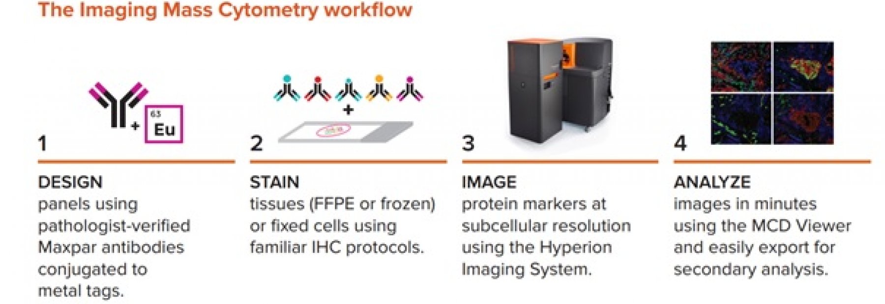
Fluidigm's hot new Hyperion system combines mass cytometry (CytoF technology) and multiparametric quantitative imaging of protein markers in tissues. The Hyperion system greatly exceeds the possibilities of classical immunohistochemical display of cell protein markers in tissues. Hyperion is not limited by optical fluorescence microscopy, but allows up to 135 detection channels. Using unique Maxpar metal antibodies, up to 40 different markers can be tracked concurrently in a single sample. In individual cells, in the context of tissue, both functional and phenotypic parameters can be simultaneously analyzed, while monitoring intracellular, extracellular and phosphorylated proteins without the risk of overlapping spectra between the detection channels.
Advantages of high-capacity protein analysis with Hyperion instrument:
- COMPLEXITY: highly multiplexed IHC, allows concurrent monitoring of up to 40 protein markers in frozen or FFPE tissues (cell surface, cytoplasm, nucleus, organelles, DNA)
- CONTEXT: maintaining cellular and tissue architecture with subcellular resolution (from 1 μm)
- EFECTIVITY: reduction of variability due to multiplex analysis of a single sample
Fluidigm Maxpar reagents
- Individual cells in the cell suspension can be labeled with more than 40 metal isotopes conjugated to Fluidim catalog antibodies or with your antibody either using a metal labeling kit or you can use the custom conjugation service Fluidigm
- Ready-to-use validated antibody-labeled panels are available against various cellular targets
Image analysis:
MCD Viewer software converts scanned cells and tissues into TIFF publication quality formats. The expression of each protein can be spectrally separated and displayed separately or in any combination with other proteins. You can cut the selected region of image (ROI) and export it to other analytics, such as histoCAT™.
System contains:
- Helios
- Hyperion
For more information see here.
PERSONAL REFERENCE
„Now we can measure so many markers simultaneously, we for the first time have the ability to determine which cell types are present in the tissue, and especially in the tumors, and also how they are spatially arranged.“
Bernd Bodenmiller, PhD Assistant Professor Institute of Molecular Life Sciences University of Zurich
Technical details about Hyperion
Web manufacturer
Go to the linkWarranty Hyperion
1 Hyperion
2 Hyperion
3 Hyperion
4 Hyperion
5 Hyperion
6 Hyperion
7 Hyperion
8 Hyperion
9 Hyperion
10 Hyperion

- Producer: Standard BioTools Inc. (Fluidigm)
Hyperion
Fluidigm's hot new Hyperion system combines mass cytometry (CytoF technology) and multiparametric quantitative imaging of protein markers in tissues. The Hyperion system greatly exceeds the possibilities of classical immunohistochemical display of cell protein markers in tissues. Hyperion is not limited by optical fluorescence microscopy, but allows up to 135 detection channels. Using unique Maxpar metal antibodies, up to 40 different markers can be tracked concurrently in a single sample. In individual cells, in the context of tissue, both functional and phenotypic parameters can be simultaneously analyzed, while monitoring intracellular, extracellular and phosphorylated proteins without the risk of overlapping spectra between the detection channels.
Advantages of high-capacity protein analysis with Hyperion instrument:
- COMPLEXITY: highly multiplexed IHC, allows concurrent monitoring of up to 40 protein markers in frozen or FFPE tissues (cell surface, cytoplasm, nucleus, organelles, DNA)
- CONTEXT: maintaining cellular and tissue architecture with subcellular resolution (from 1 μm)
- EFECTIVITY: reduction of variability due to multiplex analysis of a single sample
Fluidigm Maxpar reagents
- Individual cells in the cell suspension can be labeled with more than 40 metal isotopes conjugated to Fluidim catalog antibodies or with your antibody either using a metal labeling kit or you can use the custom conjugation service Fluidigm
- Ready-to-use validated antibody-labeled panels are available against various cellular targets
Image analysis:
MCD Viewer software converts scanned cells and tissues into TIFF publication quality formats. The expression of each protein can be spectrally separated and displayed separately or in any combination with other proteins. You can cut the selected region of image (ROI) and export it to other analytics, such as histoCAT™.
System contains:
- Helios
- Hyperion
For more information see here.
PERSONAL REFERENCE
„Now we can measure so many markers simultaneously, we for the first time have the ability to determine which cell types are present in the tissue, and especially in the tumors, and also how they are spatially arranged.“
Bernd Bodenmiller, PhD Assistant Professor Institute of Molecular Life Sciences University of Zurich
- Producer: Standard BioTools Inc. (Fluidigm)